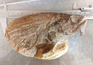
One of our 183 million year old Somerset fish fossils is on its way to be studied at the European Synchrotron Radiation Facility.
Bath Royal Literary and Scientific Institution’s Strawberry Bank Lagerstätte has been the subject of a great deal of research in recent years. One of the research tools used to study these 183 million year old fossils has been micro computed tomography (CT) scanning, using X-rays to create serial cross-sectional images. This CT scanning has enabled us to examine the internal anatomy of skeletons preserved in three dimensions, leading to fresh interpretation of organisms and understanding of their anatomy.
 During research at University of Bristol and University of Oxford, between 2016 and 2019, Dr Sam Giles discovered that some of our fish fossils from the locality have internal organs preserved, including the heart. Though the technology is impressive, the resolution of the reconstructions that can be produced using CT scanners is somewhat limited. However, Dr Giles has been granted access to a more powerful technique to study these features in this special specimen of the Lower Jurassic fish Pachycormus.
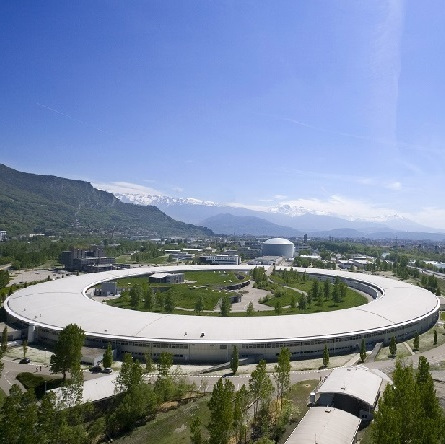
 The European Synchrotron Radiation Facility (ESRF) located in Grenoble, France, is a large particle accelerator which produces X-rays 100 billion times brighter than the X-rays used in hospitals, X-rays that allow us to fathom the structure of matter down to the minutest detail, at the atomic level. As I type Dr Giles, now Royal Society Dorothy Hodgkin Research Fellow at the University of Birmingham
and Academic Keeper of the Lapworth Museum of Geology, is traveling to Grenoble to reveal the internal anatomy of this fish.
You can read more about Strawberry Bank here and find out about the ESRF here.